Negative Pressure Wound Therapy (NPWT) for Infected Wounds: A Complete Guide

Introduction

Infected wounds present a significant challenge in wound care, requiring specialized treatment to prevent complications and promote healing. Negative Pressure Wound Therapy (NPWT) has proven effective in managing and treating infected wounds by creating an optimal environment for recovery.

How NPWT Helps in Infected Wound Management?

  1. Reducing Bacterial Load: Continuous suction removes fluids containing bacteria and necrotic debris, minimizing the risk of further infection.
  2. Enhancing Blood Circulation: Promotes oxygen and nutrient delivery to the wound bed, accelerating healing.
  3. Controlling Exudate and Moisture: Prevents excessive fluid buildup, which can contribute to bacterial growth.
  4. Encouraging Tissue Regeneration: Stimulates granulation tissue formation, helping wounds heal faster.
  5. Reduce Inflammatory Response: Minimizes swelling and pain.
  6. Support Antibiotic Therapy: Complements systemic treatments by maintaining a clean wound bed.
  7. Reducing the Need for Frequent Dressing Changes: Minimizes exposure to external contaminants and enhances patient comfort.

Benefits of NPWT for Infected Wounds

  1. Accelerated Healing: NPWT creates a controlled environment for rapid wound closure 
  2. Infection Control: NPWT helps prevent sepsis and systemic infections.
  3. Reduced Amputation Risk: NPWT minimizes the likelihood of limb amputation.
  4. Cost-Effectiveness: NPWT decreases overall treatment costs by reducing hospital stays and complications.
  5. Versatility: Effective for various wound types, including post-surgical infections, traumatic injuries, and chronic ulcers.

When is NPWT Recommended for Infected Wounds?

  1. Post-Surgical Infections
    • Examples: Dehisced wounds or abscesses post-abdominal or orthopedic surgeries.
    • Benefit: Prevents deep tissue infections and promotes closure.
  2. Traumatic Wounds
    • Examples: Contaminated lacerations or crush injuries.
    • Benefit: Removes debris and maintains clean wound conditions.
  3. Diabetic Foot Ulcers
    • Problem: High risk of infection and delayed healing.
    • Solution: NPWT minimizes bacterial load and supports tissue repair.
  4. Chronic Wounds
    • Examples: Pressure ulcers and venous leg ulcers.
    • Benefit: Accelerates granulation and manages recurring infections.
  5. Burn Wounds
    • Problem: High susceptibility to infection.
    • Solution: NPWT provides a sterile environment and prevents sepsis.

Step-by-Step Process of Using NPWT for Infected Wounds

  1. Wound Assessment
    • Clean and debride necrotic tissue.
    • Evaluate wound depth and infection severity.
  2. Dressing Application
    • Place foam or gauze dressing tailored to the wound shape.
  3. Sealing
    • Apply an airtight drape to isolate the wound.
  4. Vacuum Activation
    • Connect the dressing to a pump and set appropriate negative pressure.
    • Continuous or intermittent suction is used depending on wound characteristics.
  5. Monitoring and Maintenance
    • Replace dressings every 48–72 hours.
    • Monitor for infection signs like fever, redness, or odor.

Risks and Contraindications for NPWT in Infected Wounds

Key Risks:

  1. Pain: Negative pressure may cause discomfort, especially during dressing changes.
  2. Bleeding: Fragile wounds may bleed if pressure settings are too high.
  3. Device Malfunctions: Ensure the pump and tubing are functioning correctly.

Contraindications:

  1. Avoid untreated osteomyelitis or wounds with excessive necrotic tissue.
  2. Not suitable for patients with uncontrolled bleeding disorders.

Practical Tips for Effective NPWT Use in Infected Wounds

  1. Combine with Antibiotics
    • Use NPWT alongside systemic antibiotics for effective infection management.
  2. Regular Dressing Changes
    • Adhere to dressing schedules to prevent bacterial growth.
  3. Patient Education
    • Train patients on device care if used at home.
  4. Monitor Progress
    • Regularly evaluate wound size, exudate levels, and infection markers.

Clinical Evidence Supporting NPWT for Infected Wounds

  •  A 2018 study in the International Wound Journal reported a significant reduction in bacterial load in NPWT-treated wounds.
  • A 2014 Open Orthopaedics Journal study found that NPWT significantly lowered infection rates in high-risk traumatic wounds, especially when combined with surgical debridement.

Frequently Asked Questions (FAQs)

1. What are the signs that my infected wound is not improving with NPWT?

If you notice increased redness, swelling, warmth, pain, a foul odor, or pus-like drainage, it could indicate a worsening infection. Contact your healthcare provider immediately.

NPWT helps manage infection by removing bacteria and promoting healing, but it is not a substitute for antibiotics or surgical debridement when necessary. Your doctor will determine the best combination of treatments.

Dressing changes may be needed more frequently for infected wounds, sometimes every 24-48 hours, depending on the level of drainage and infection severity. Your provider will decide on the appropriate schedule.

If used improperly or if the infection is severe, bacteria can be spread. That’s why NPWT for infected wounds should always be managed by a trained healthcare professional.

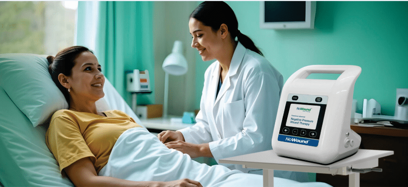
Check the device screen for alerts. Common issues include loss of suction, tubing blockages, or full canisters. Contact your healthcare provider immediately if you’re unsure how to fix it.

Some discomfort may occur, especially during dressing changes. To minimize discomfort, your provider may recommend pain relief before dressing changes.

Final Thoughts: Why NPWT is Right for Your Infected Wound

NPWT can provide significant benefits, particularly for slow-healing, infected wounds. By reducing infection risks and promoting faster recovery it offers hope for a better quality of life. Consult your doctor to determine if NPWT is the right solution for managing your wound.

24/7 Patient Helpline

9773941323

References

  1. https://onlinelibrary.wiley.com/doi/abs/10.1111/
  2. https://openorthopaedicsjournal.com/VOLUME/8/PAGE/168/FULLTEXT/